Magnetic Resonance Imaging
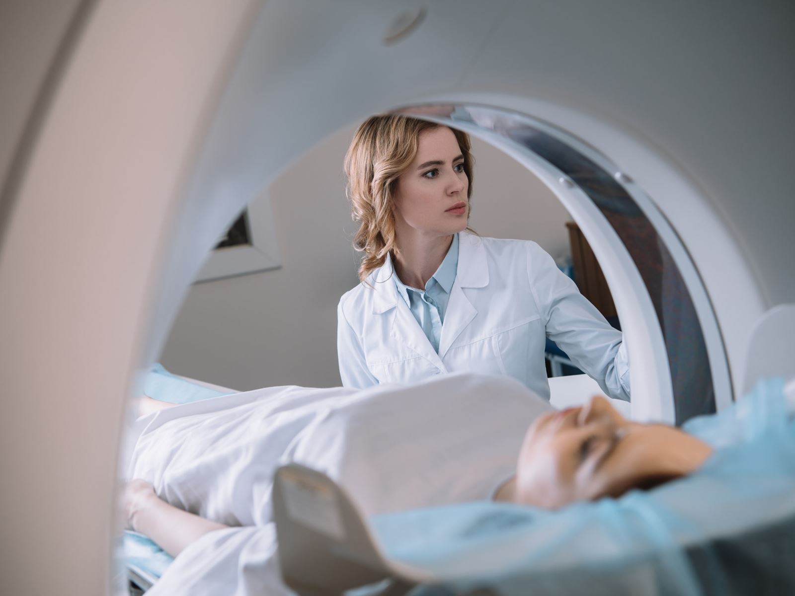
Our Magnetic Resonance Imaging (MRI) program is designed with the patient in mind. The scanning center is located on the second lower level (L2) of the Hale Building for Transformative Medicine (60 Fenwood Road) at Brigham and Women’s Hospital. This location provides patients with the convenience of having their scans done in the same building as the MS Center and infusion suite. The MRI center features state-of-the-art imaging equipment including a 3T and 7T magnet. Patients receive their routine imaging on the 3T scanner, which provides excellent pictures allowing the tracking of disease activity and monitoring for disease progression by the detection of lesions and atrophy. The suite also features a 7T magnet, which provides a unique opportunity for our patients – this is one of the few 7T scanners in the United States that is approved for routine care. It may be recommended to patients to get a brain scan on the 7T device to complement the information obtained at 3T. The 7T brings the benefit of a more accurate diagnosis in uncertain cases by the detection of the central vein sign to help determine if the patient’s lesions are due to MS. The 7T also has unique sensitivity to gray matter lesions both in the cerebral cortex and deep gray matter. In addition, with contrast infusion (IV gadolinium), the 7T can investigate whether patients have involvement of the covering of the brain (the leptomeninges). The 7T MRI unit is a unique imaging device that has already had an immediate impact on our research, and may eventually help improve the care of patients via its ability to produce exceptionally high-quality images.