
Neuromyelitis Optica
Neuromyelitis optica (NMO) is a disease of the central nervous system that typically affects vision and walking by attacking the optic nerves and spinal cord. Although NMO can appear similar to multiple sclerosis, it can also cause a variety of other symptoms and is a different condition requiring its own specialized treatment.
At the Brigham Multiple Sclerosis Center, NMO experts manage complex care for patients while engaging in research to better understand its behavior and one day, its cause. In addition to our core team, care is also available through our relationships with world-class colleagues in physical therapy, neuro-ophthalmology, urology, psychiatry, and social work.
Our team has been involved in projects to understand:
- Which treatments are most effective in managing NMO
- How to distinguish NMO not only from MS, but also other related rare disorders
- Differences in the behavior of NMO in children compared to adults
- How imaging and biomarker tools help assess disease activity and aid treatment decisions
- How pregnancy and hormonal changes influence NMO, and vice versa
We are engaged in both local and international collaborations with the goal of identifying, understanding, and ultimately defeating this rare but serious condition.
More Information
Signs & Symptoms
NMO symptoms can vary from person to person and may resemble MS symptoms in many ways. The most common symptoms are:
- Optic neuritis
- Transverse myelitis
- Shooting pain or tingling in the neck, back or abdomen
- Loss of bowel and bladder control
Optic neuritis is inflammation of the optic nerve that causes pain with eye movement and decreased vision in the affected eye. Transverse myelitis is inflammation of the spinal cord that may cause limb weakness, numbness or partial paralysis.
Although rare, symptoms outside of the optic nerves and spinal cord do occur, such as uncontrollable vomiting and hiccups, which are now being recognized as symptoms of NMO or NMOSD due to brainstem involvement.
In the past, NMO was diagnosed in patients who experienced a rapid onset of blindness in one or both eyes, followed within days or weeks by varying degrees of paralysis in the arms or legs. In most cases, however, the interval between optic neuritis and transverse myelitis is significantly longer, sometimes as long as several years. After the initial attack, NMO follows an unpredictable course.
During a relapse, new damage to the optic nerves and/or spinal cord can lead to accumulating disability; therefore preventing attacks is critical to a good long-term outcome. Sometimes these symptoms are temporary, and resolve on their own. In any case, it is important to discuss these symptoms with your doctor to help consider NMO in your diagnosis.
Treatment
There is no cure for NMO at this time, and no medications are specifically approved to treat it. The standard of care for an initial attack of NMO includes the following:
- Intravenous high-dose corticosteroids (methylprednisolone)
- Plasma Exchange (PLEX) if no improvement occurs with corticosteroids. The goal of PLEX is to lower the level of NMO-IgG in the blood. PLEX involves removing blood from the body through a needle and tubing. Through a series of steps, the plasma (the liquid part of the blood) is separated from blood cells and replaced with an artificial plasma substitute; the plasma substitute and blood cells are combined and returned to the body through an intravenous line. The procedure lasts several hours and may be repeated multiple times over a number of days.
Because the likelihood of recurrence is greater than 90 percent and attacks are generally severe, ongoing treatment to suppress the immune system is considered necessary. Some of the medications used for maintenance therapy include:
- eculizumab (Solaris)
- inebilizumab (Uplizna)
- satralizumab (Enspryng)
Read more about the benefits and risks of these medications. The Brigham MS Center also outlines some of these treatments here.
NMO Research
- Female hormonal exposures and neuromyelitis optica symptom onset in a multicenter study.
Bove R, Elsone L, Alvarez E, Borisow N, Cortez MM, Mateen FJ, Mealy MA, Mutch K, Tobyne S, Ruprecht K, Buckle G, Levy M, Wingerchuk DM, Paul F, Cross AH, Weinshenker B, Jacob A, Klawiter EC, Chitnis T. - Child Neurology: Neuromyelitis optica spectrum disorders.
Bradshaw MJ, Vu N, Hunley TE, Chitnis T. Neurology. 2017 Jan 10;88(2):e10-e13. doi: 10.1212/WNL. 0000000000003495. No abstract available. PMID: 28069981 - Immunology of neuromyelitis optica during pregnancy.
Davoudi V, Keyhanian K, Bove RM, Chitnis T. Immunology of neuromyelitis optica during pregnancy. Neurol Neuroimmunol Neuroinflamm. 2016 Oct 7;3(6):e288. Review. PubMed PMID: 27761482; PubMed Central PMCID: PMC5056648. - Neuromyelitis optica spectrum disorders in children and adolescents.
Tenembaum S, Chitnis T, Nakashima I, Collongues N, McKeon A, Levy M, Rostasy K. Neuromyelitis optica spectrum disorders in children and adolescents. Neurology. 2016 Aug 30;87(9 Suppl 2):S59-66. PubMed PMID: 27572863. - Clinical features of neuromyelitis optica in children: US Network of Pediatric MS Centers report.
Chitnis T, Ness J, Krupp L, Waubant E, Hunt T, Olsen CS, Rodriguez M, Lotze T, Gorman M, Benson L, Belman A, Weinstock-Guttman B, Aaen G, Graves J, Patterson M, Rose JW, Casper TC. Clinical features of neuromyelitis optica in children: US Network of Pediatric MS Centers report. Neurology. 2016 Jan 19;86(3):245-52. PubMed PMID: 26683648; PubMed Central PMCID: PMC4733158. - Update on biomarkers in neuromyelitis optica.
Melamed E, Levy M, Waters PJ, Sato DK, Bennett JL, John GR, Hooper DC, Saiz A, Bar-Or A, Kim HJ, Pandit L, Leite MI, Asgari N, Kissani N, Hintzen R, Marignier R, Jarius S, Marcelletti J, Smith TJ, Yeaman MR, Han MH, Aktas O, Apiwattanakul M, Banwell B, Bichuetti D, Broadley S, Cabre P, Chitnis T, De Seze J, Fujihara K, Greenberg B, Hellwig K, Iorio R, Jarius S, Klawiter E, Kleiter I, Lana-Peixoto M, Nakashima, O’Connor K, Palace J, Paul F, Prayoonwiwat N, Ruprecht K, Stuve O,Tedder T, Tenembaum S, Garrahan JP, Aires B, van Herle K, van Pelt D, Villoslada P, Waubant E, Weinshenker B, Wingerchuk D, Würfel J, Zamvil S. Update on biomarkers in neuromyelitis optica. Neurol Neuroimmunol Neuroinflamm. 2015 Jul 23;2(4):e134. Review. PubMed PMID: 26236760; PubMed Central PMCID: PMC4516398. - Protective environmental factors for neuromyelitis optica.
Graves J, Grandhe S, Weinfurtner K, Krupp L, Belman A, Chitnis T, Ness J, Weinstock-Guttman B, Gorman M, Patterson M, Rodriguez M, Lotze T, Aaen G, Mowry EM, Rose JW, Simmons T, Casper TC, James J, Waubant E; US Network of Pediatric Multiple Sclerosis Centers. Protective environmental factors for neuromyelitis optica. Neurology. 2014 Nov 18;83(21):1923-9. PubMed PMID: 25339213; PubMed Central PMCID: PMC4248458. - Predictors of recurrence following an initial episode of transverse myelitis.
Kimbrough DJ, Mealy MA, Simpson A, Levy M. Predictors of recurrence following an initial episode of transverse myelitis. Neurol Neuroimmunol Neuroinflamm. 2014 Apr 24;1(1):e4. PubMed PMID: 25340060; PubMed Central PMCID: PMC4202674. - International consensus diagnostic criteria for neuromyelitis optica spectrum disorders.
Wingerchuk DM, Banwell B, Bennett JL, Cabre P, Carroll W, Chitnis T, de Seze J, Fujihara K, Greenberg B, Jacob A, Jarius S, Lana-Peixoto M, Levy M, Simon JH, Tenembaum S, Traboulsee AL, Waters P, Wellik KE, Weinshenker BG; International Panel for NMO Diagnosis.. International consensus diagnostic criteria for neuromyelitis optica spectrum disorders. Neurology. 2015 Jul 14;85(2):177-89. PubMed PMID: 26092914; PubMed Central PMCID: PMC4515040. - Demographic and clinical features of neuromyelitis optica: A review.
Pandit L, Asgari N, Apiwattanakul M, Palace J, Paul F, Leite MI, Kleiter I, Chitnis T; GJCF International Clinical Consortium & Biorepository for Neuromyelitis Optica. Demographic and clinical features of neuromyelitis optics: A review. Mult Scler. 2015 Jun;21(7):845-53. Review. PubMed PMID: 25921037; PubMed Central PMCID: PMC4463026. - Treatment of Neuromyelitis Optica: Review and Recommendations.
Kimbrough DJ, Fujihara K, Jacob A, Lana-Peixoto MA, Leite MI, Levy M, Marignier R, Nakashima I, Palace J, de Seze J, Stuve O, Tenembaum SN, Traboulsee A, Waubant E, Weinshenker BG, Wingerchuk DM; GJCF-CC&BR. Treatment of Neuromyelitis Optica: Review and Recommendations. Mult Scler Relat Disord. 2012 Oct;1(4):180-187. PubMed PMID: 24555176; PubMed Central PMCID: PMC3926208.
Diagnosis
MRI
With so many symptoms in common, NMO can sometimes be confused with MS or other diseases that are treated in different ways. Early detection is crucial for the best outcome. MRI has been an extremely valuable tool used to establish an early diagnosis and monitor disease progression in NMO. Click here to download and read GJCF's helpful and informational NMO Patient Resource Guide. On pages 30 and 31, the resource guide states that "Generally MRI test results in NMO patients show lesions indicative of inflammation that extend over three or more segments of the spinal cord, the optic nerve(s) and occasionally in the brain. However, brain lesions observed in NMO follow a different pattern and are not as common as in other diseases, including MS." This is one example of how physicians used MRI to rule out other diagnoses, especially MS.
Blood Test – NMO-IgG
As mentioned previously, in the blood of NMO patients a particular antibody is present, AQP4-IgG. The blood test that any physician can order is known as NMO-IgG. Its detection strongly supports a diagnosis of NMO, as approximately 75% of patients with NMO have this antibody in their blood serum and it is not found in people with MS or other conditions. However, the spectrum of NMO disorders also includes patients whose antibody status is unknown or negative but still have the core clinical features and neuroimaging findings, and therefore, a postive NMO-IgG is not absolutely necessary to make the diagnosis of NMOSD. Additionally, some patients may have undetectable antibody levels due to treatment they are receiving and retesting may be necessary. Newer tests are being developed that have a higher rate of detecting the NMO-IgG antibody.
Lumbar Puncture
If the blood test for NMO-IgG is negative and the diagnosis is unclear, a neurologist may suggest a lumbar puncture or "spinal tap" for further testing. The lumbar puncture allows the neurological team to test the cerebrospinal fluid (CSF) surrounding the brain and spinal cord, and observe the levels of immune cells, proteins and antibodies in the fluid. In NMO, the spinal fluid may show considerably elevated white blood cells during attacks, which are greater than typically seen in other autoimmune diseases. In differentiating these conditions CSF is tested for oligoclonal bands (proteins), which are commonly detected in MS patients, but usually (not always), negative in patients with NMO.
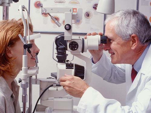
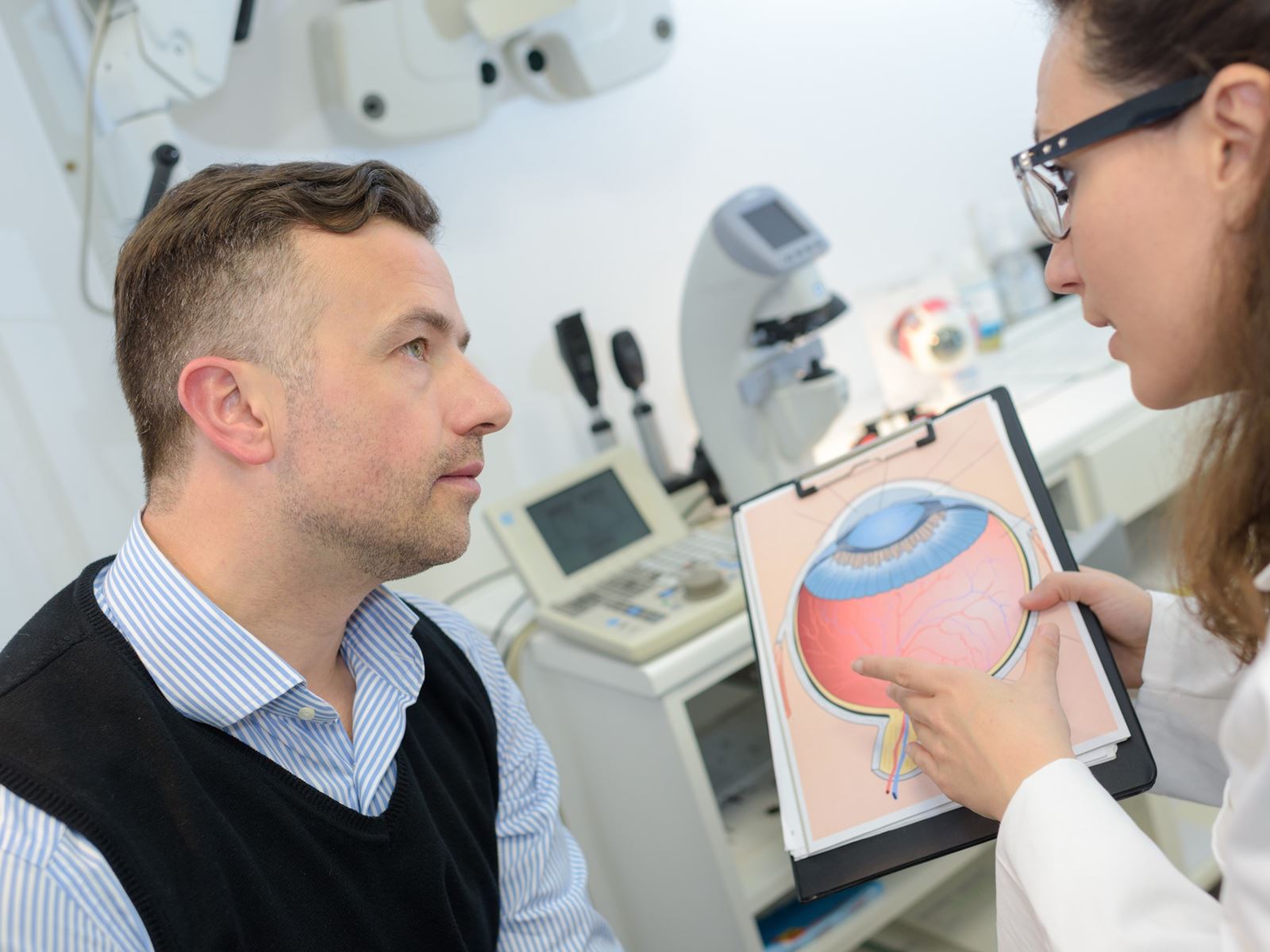
Ophthalmological Tests
To help obtain a correct diagnosis, patients may be referred to an ophthalmologist or eye specialist. The ophthalmologist may perform the following eye tests:
- A routine eye exam will check vision and the ability to perceive different colors and depth perception.
- Ophthalmoscopy examines the structures at the back of the eye by shining a bright light into the area. This eye test evaluates the optic disk, which is the area where the optic nerve enters the retina in the eye. The optic disk becomes temporarily swollen in about one-third of people with optic neuritis (ON). Patients who have had previous optic neuritis due to NMO may have a permanently pale optic disk, but the same may occur in patients with MS and other conditions that target the optic nerve, therefore this is not a specific finding for NMO.
- Pupillary light reaction tests the eyes to see how pupils respond when exposed to bright light. After shining a bright light in a healthy eye, the pupil of the eye affected by ON often incorrectly dilates.
- Optical coherence tomography (OCT) is a non-invasive image technique to study the retina. OCT is a simple high-resolution scan used to measure the thickness of the retinal nerve fiber layer (RNFL). The RNFL is often decreased in NMO patients with optic neuritis.