

About MS
Multiple sclerosis (MS), originally described in the 1800s, is a chronic neurological disease affecting the central nervous system. The central nervous system includes the brain and spinal cord. Multiple Sclerosis is an autoimmune disease, which means that our immune system is activated to react against our own nervous system. In multiple sclerosis, the main target of this immune attack is the myelin covering over an axon. The axons are part of a neuron (nerve cell) and are used to transmit information to other nerve cells. Efficient transmission is maintained with the presence of myelin, analogous to rubber insulation over an electrical wire. Neurological signs typical for multiple sclerosis occur when there has been myelin breakdown.


MS Patterns
There are typical patterns for the disease course and patients can be categorized within one of these groups:
- Relapse: A new neurological deficit which lasts for more than 24 hours. Attack or exacerbation are other names for a relapse.
- Pseudorelapse (Pseudoexacerbation): Occasionally, pre-existing symptoms may worsen, mimicking a relapse. This is usually associated with the onset of a fever, infection, or extreme fatigue. The treatment for a pseudorelapse is treatment of the underlying infection.
- Secondary Progressive MS: Is a form of MS that generally occurs in relapsing-remitting patients 10-15 years into the course of disease. In this form of MS, neurological deficits do not remit, but persist and cause chronic symptoms.
- Primary Progressive MS: Approximately 10% of patients experience primary progressive MS, in which progression occurs from the onset of disease. Primary progressive MS often begins after age 40 years, which differs from the typical age of onset of RRMS (20s-30s). An increased proportion of men experience primary progressive MS compared to other forms of MS.
- Progressive relapsing MS: A minority of patients experience a course of disease in which relapses and progression are superimposed from the onset of disease.
- Neuromyelitis Optica (NMO): A rare autoimmune disorder that affects the optic nerves and spinal cord almost exclusively. An antibody marker in the serum is present in approximately 75% of patients. Click here to learn more about NMO.

Figure 1
MS World Distribution
The prevalence of MS is highest in North America, northern Europe and southern Australia. There is a complex pattern of prevalence within these areas (figure 1). In North America, the highest rate is in southern Canada and in the northern United States. The lowest rate in North America is the Mississippi region and surrounding southeastern states. Studies have indicated that early life residence influences the risk of developing multiple sclerosis. These findings suggest environmental factors for the development of the disease, though nothing has been isolated. Additional evidence suggests genetic factors to be involved. Racial susceptibility studies have found Scandinavian descent populations are at a higher risk compared to people of African descent. There has been a genetic marker found in select populations with MS but the mode of transmission and the influence of additional genes is not understood. The genetic susceptibly of MS is the focus of abundant research. Overall, there appears to be combination of genetic and environmental factors contributing to the disease.
MS Distribution
Multiple Sclerosis (MS) is considered to be an immune mediated disease. Typically, when our body's immune response is triggered, lymphocytes including T cells and B cells will be activated. Depending on the type of defense our body requires; various subsets of cells will be activated. In MS, T cells become activated in a genetically susceptible person by a probable environment factor and migrate to the central nervous system (figures 2 & 3). During this activation, cytokines are released to facilitate activation and to enhance the ability of T-cells to travel into the central nervous system (figure 6). Modifications of cytokines are heavily studied in new therapeutic strategies for MS. There are cells called macrophages and microglia that bind antigens, the target for the immune attack. These macrophages and microglia are called antigen-presenting cells. These cells form a complex with the activated T cells (figure 4). This triggers a cascade of events which ultimate leads to the destruction of the myelin and oligodendrocytes, which are cells that produce myelin (figure 5 & figure 6). There has been a significant amount of research indicating destruction of axons is occurring during this process. Areas of the central nervous system affected are referred to as "lesions" or "plaques" (figure 7). Different pathological patterns of demyelination and axonal loss have been discovered in lesions from brains of patients with MS.
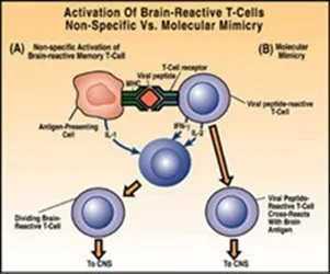
Figure 2
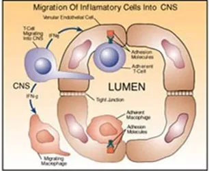
Figure 3
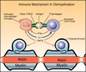
Figure 4
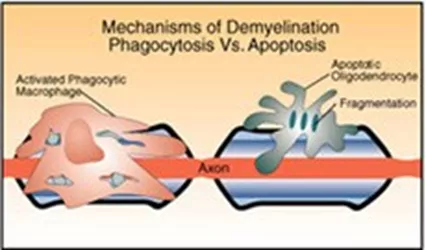
Figure 5
Neuromyelitis Optica
Neuromyelitis optica (NMO) is a disease of the central nervous system that typically affects vision and walking by attacking the optic nerves and spinal cord. Although NMO can appear similar to multiple sclerosis, it can also cause a variety of other symptoms and is a different condition requiring its own specialized treatment.
At Mass General Brigham/MGB, NMO experts manage complex care for patients while engaging in research to better understand its behavior and one day, its cause. In addition to our core team, care is also available through our relationships with world-class colleagues in physical therapy, neuro-ophthalmology, urology, psychiatry, and social work.
Our team has been involved in projects to understand:
- Which treatments are most effective in managing NMO
- How to distinguish NMO not only from MS, but also other related rare disorders
- Differences in the behavior of NMO in children compared to adults
- How imaging and biomarker tools help assess disease activity and aid treatment decisions
- How pregnancy and hormonal changes influence NMO, and vice versa
We are engaged in both local and international collaborations with the goal of identifying, understanding, and ultimately defeating this rare but serious condition.
Patient Resources
The National MS Society has brochures, booklets, and books about many aspects of MS. For example, they have booklets directed at children of parents with MS, teenagers who have a parent with MS, and another one about working together as a family. Many of their print resources are available on their web site, and are also available at your local chapter. You can phone their central number and be connected to your local chapter: 1-800-Fight-MS (1-800-344-4867)
Dr. Howard Weiner, the Co-Director of Partners MS Center in Boston, has written a new book called Curing MS: How Science is Solving the Mysteries of Multiple Sclerosis, 2004, Random House. It is a very interesting book, which takes you behind the scenes in the labs as scientists and doctors study and test treatments for MS. It can help you gain a solid understanding of MS without being a list of all the bad things that the illness might do to you.
Roz Kalb is a psychologist who has worked for many years with people with MS and their families. She has authored many books and articles. Here are three suggested readings:
- Kalb, Rosalind,Ed., (2000): Multiple Sclerosis: the questions you have, the answers you need, (2nd Edition), Demos Publications, 386 Park Avenue South, Suite 201, New York, NY 10016, U.S.A.
- Kalb, Rosalind,Ed., (1998): Multiple Sclerosis: A Guide for Families. New York: Demos Publications.
- LaRocca, Nicholas, & Kalb, Rosilind (2006): Mult
Websites
- www.nmss.org is the website of the National Multiple Sclerosis Society, and it is full of useful information. The MS Society is fairly conservative about the information published, so it is very reliable.
- www.msif.org is the website of the Multiple sclerosis international federation. Their website is a link to people with MS all over the world, and it is a good site to learn about current research. They send out an email newsletter which updates people about current published research.
- www.ninds.nih.gov/disorders/multiple_sclerosis/multiple_sclerosis.htm is the website of the National Institute of Neurological Disorders and Stroke, of the National Institutes of Health. This is the government agency which funds research into MS, and the information on this website is reliable.
- www.acceleratedcure.org is the website of the Accelerated Cure Project, a new organization seeking to enhance MS research.
- www.mscare.org is the website of the Consortium of Multiple Sclerosis Centers, a national organization which is a link between MS Centers, and a good source of reliable information.
- www.expertmspatient.com is an educational tool offered by the Consortium of MS Centers
- www.partnerspediatricmscenter.org is the website of the Partners Pediatric MS Center at Massachusetts General Hospital for Children.
- www.nationalmssociety.org/keepsmyelin/index.html is the website of the online magazine, "Keep Smyelin", an entertaining publication for children who have a parent or relative with MS.
- www.lookingglass.org is the website of Through the Looking Glass, an organization in California which advocates for people with disabilities and in particular for parents with a disability.
- www.sexualhealth.com A website that includes an article about MS and sexuality in its Disability and Chronic Illness section.
- Each of the companies that make medications for MS, have their own websites. These sites often have useful information directed at patients who are using their medication. They are also commercial sites that are used to market each company’s medication. Patients find these sites useful, but remember to read the information in the light of the marketing efforts employed there.
Financial Assistance Resources
- Adapting: Financial Planning for a Life with Multiple Sclerosis, A free workbook offered by the National MS Society to help you manage your money and plan wisely for the future.
- Home LINKS Program 800-493-9255 or www.nationalmssociety.org/MAM/event/event_detail.asp?e=13699
Offered by the New England Chapter of the MS Society, the Home LINKS program is a short-term care management program providing information and referral, support, access to care, individualized care management and self-advocacy tools. - www.ilusa.com Independent Living Centers are typically non-residential, private, non-profit, consumer-controlled, community-based organizations providing services and advocacy by and for persons with all types of disabilities. Their goal is to assist individuals with disabilities to achieve their maximum potential within their families and communities. This website provides a listing of all the Independent Living Centers in your state.
- MassHealth/Medicaid 800-841-2900
- Medicare 800-633-4227
Massachusetts Resources
Day Treatment
- B.Fit 617-825-3905×300 or www.thebostonhome.org an innovative, outpatient day program for adults with multiple sclerosis and other neurological diseases.
Employment/Disability
- Mass Rehab Commission 1-800-223-2559 or www.mass.gov
- Resource Partnership 508-647-1722 or www.resourcepartnership.org
- Social Security Administration 800-772-1213 www.ssa.gov.
- Information about eligibility for SSI and SSDI. Applications can be made on-line.
Financial Assistance
- www.MassResources.org provides detailed information about the many types of assistance available to those in need in Massachusetts including housing, food, utility
assistance, legal assistance, etc.
Transportation
- "The Ride" 617-222-5123 or www.mbta.com
- Download the application for The Ride, which provides door-to-door transportation for people who cannot use general public transportation.
New Hampshire Resources
- New Hampshire Helpline 603-225-9000 or by dialing 2-1-1 from an in state phone
- or www.nhhelpline.org Information on social services and emergency help.
Rhode Island Resources
- MS Society Rhode Island Chapter 401-738-8383
- Office of Community Services & Advocacy 401-421-7833.
- Under the auspices of Catholic Diocese of Providence, they operate several centers that provide an array of community based services including homebound care and respite care services.