Bakshi Lab: Laboratory for Neuroimaging Research
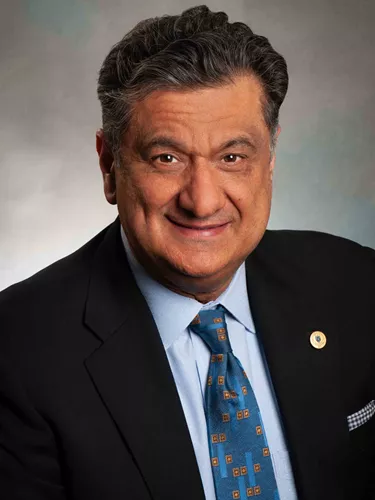
Under the direction of Dr. Rohit Bakshi, the Laboratory for Neuroimaging Research (LNR) continues to refine technical aspects of MRI imaging in order to better understand the pathophysiology of MS. Seminal work has been conducted on brain atrophy including gray matter atrophy, spinal cord involvement, and the role of neurodegeneration in MS.
The publications from the LNR are listed here.
Center for Neurological Imaging
The MRI program at the Brigham MS Center is directed by Dr. Rohit Bakshi. This entails several key aspects of understanding how to use MRI in studying multiple sclerosis. From 2000 to 2014, the MS Center performed routine scans on a 1.5T scanner. We then moved to a 3T scanner giving us better pictures of the brain and spinal cord. Most recently, in 2017, the hospital installed a 7T scanner, bringing our capability to image the brain to a high level of detail for our research. In 2018, the 7T scanner was approved by the FDA and Massachusetts for routine patient care. Patients currently receive scans at both 3T and 7T to provide the most complete information on the status of their MS. The 7T scanner is particularly helpful to define central veins within lesions, and MS involvement of the gray matter and the covering of the brain (the leptomeninges). The Center has advanced tools to process and analyze MRI scans including a database management operation to store and organize all MRI scans obtained. This currently holds more than 25,000 unique MRI scans obtained since the year 2000 in the CLIMB study. We have developed state-of-the-art methods to analyze our scans such as 3T morphometry.


